Postări

The problem of the HINGE axis concept!

Imagine
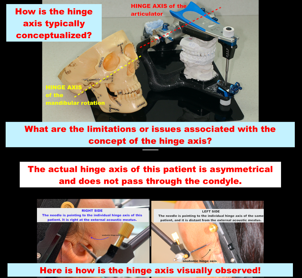
Let’s start with the concept of the HINGE AXIS. We have been taught that the hinge axis is located somewhere in the condyle. If the hinge axis is indeed located within the condyle, then human beings invented the articulator. The articulator replicates the movements and the position of the lower jaw relative to the skull! Working with the articulator is akin to working directly on the human being, given that it replicates the same hinge axis recorded from the patient. After the recording of the hinge axis, we visualize it as depicted in the picture. ONE PROBLEM! Recording the hinge axis on condylography precisely determines the hinge axis. Condylography is a computerized method for determining the hinge axis and tracking the movements of the jaws. After removing the sensors, we can accurately mark the exact location of the hinge axis. CONCLUSION FOR THE RIGHT JOIN: The hinge axis is located at the margin of the external ear duct. While we were taught that the anatomical hinge axis is ty

The HINGE AXIS concept!

Imagine
How do we conceptualize the hinge axis and how does it function in practice? The hinge axis of mandible rotation, also known as the mandibular hinge axis, is an anatomical reference point representing the axis around which the lower jaw or mandible can rotate during its movements. This axis is crucial in the field of dentistry and maxillofacial sciences for various diagnostic and treatment purposes, particularly in relation to the temporomandibular joint (TMJ) and occlusion studies. The actual hinge axis can be determined through three methods: 1. Anatomic 2. Graphic/needle and paper/ 3. Computerized/electronic (CADIAX) The last method is considered the most precise. THE RESULT OF INVESTIGATION OF THE COMPUTERIZED/electronic(CADIAX)/ The needle is pointing to the individual hinge axis of this patient. It is right at the external acoustic meatus. RIGHT SIDE LEFT SIDE CONCLUSION: The hinge axis of this patient is not within the cond yle, and it is asymmetrical. ONE MORE QUESTION! How can

THE HINGE AXIS CONCEPT

Imagine
  What types of hinge axis do we have? #terminal hinge position #hinge movement #hinge axis The terminal hinge position occurs when the condyles are located in the articular fossae, and the mandible is capable of pure rotary opening. In Centric Relation (CR), the mandible can rotate around the horizontal axis for approximately 20-25 mm, and this measurement is typically taken between the maxillary and mandibular incisal edges of the teeth. The horizontal axis around which the hinge movement occurs is referred to as the hinge axis. Since the temporomandibular joint (TMJ) has two condyles, the right condyle connects imaginarily with the right condyle to create an axis of rotation.”     HOW DO WE RECORD THE HIINGE AXIS? I am utilizing the KINEMATIC FACEBOW technology, which allows me to assess the following information: • Anatomic hinge axis • Graphic/needle and papaer • Computerized  hinge axis THE ANATOMIC HINGE AXIS “It is considered that the anatomic hinge axis is around 11-

AnteriorUnforcedPosition

Imagine
               My question about the AnteriorUnforcedPosition is:                                                   How does the patient brux in this condylar position?       Here is a case presentation about the bruxism of the patients in the anterior unforced position. Step 1: The anterior unforced position was determined using the Gothic arch. Step 2. The anterior unforced position was recorded. Step 3: A splint was fabricated in this position. Step 4: Bruxism was assessed using a bruxchecker. The initial recording was taken before testing with the bruxchecker. The second recording was obtained after testing with the bruxchecker. Step 5: The results indicate that the patient remains very calm in the anterior unforced position.

TP1

Imagine
The diagram below shows the protrusion and open/close movement of the condylar hinge axis.  When both lines overlap, it indicates that the recording was done correctly.  The therapeutic position was recorded on the Gothic Arch. As you can see, the TP is not in front of the arrow, but somewhere in the middle. I performed the condylography measurement and found that the therapeutic position is at approximately 2.74 mm on the right side and 1.24 mm on the left side from the reference position (RP).  Please check the condylographic tracings.   The Gothic arch was designed on these tracings, and the therapeutic position was fixed based on these designs. The results of the condylar reposition are shown in the pictures below! Right side Before condylar reposition After condylar repositioning                                                                            Left side  Before condylar repositioning After condylar repositioning